Retinal Vein Occlusion
Retinal Vein Occlusion (RVO) occurs when one of the veins that carries blood to the retina becomes blocked, causing blood and fluid to leak into the retinal tissue. This causes blurred vision and is often compared to a stroke occurring within the eye. There are two main types: Central Retinal Vein Occlusion (CRVO), which affects the eye’s main retinal vein and can cause significant vision loss, and Branch Retinal Vein Occlusion (BRVO), which involves a smaller vein and usually results in more localised damage.
Symptoms and Risk Factors
RVO can develop due to factors such as high blood pressure, glaucoma, diabetes, high cholesterol, smoking, blood-clotting disorders, or dehydration. Common symptoms include blurred vision and floaters. Pain is uncommon unless complications develop later. The risk of retinal vein occlusion increases with age, particularly in people over 50.
Types of Retinal Vein Occlusion
Central retinal vein occlusion affects the main retinal vein and often causes widespread retinal swelling and vision loss. Branch retinal vein occlusion occurs when a smaller branch vein is blocked, usually affecting a specific area of the retina. Vision loss may be milder in branch retinal vein occlusion but can still be significant if the macula is involved. Identifying the type of retinal vein occlusion helps guide appropriate treatment and monitoring.
Both types require specialist assessment to reduce the risk of complications.
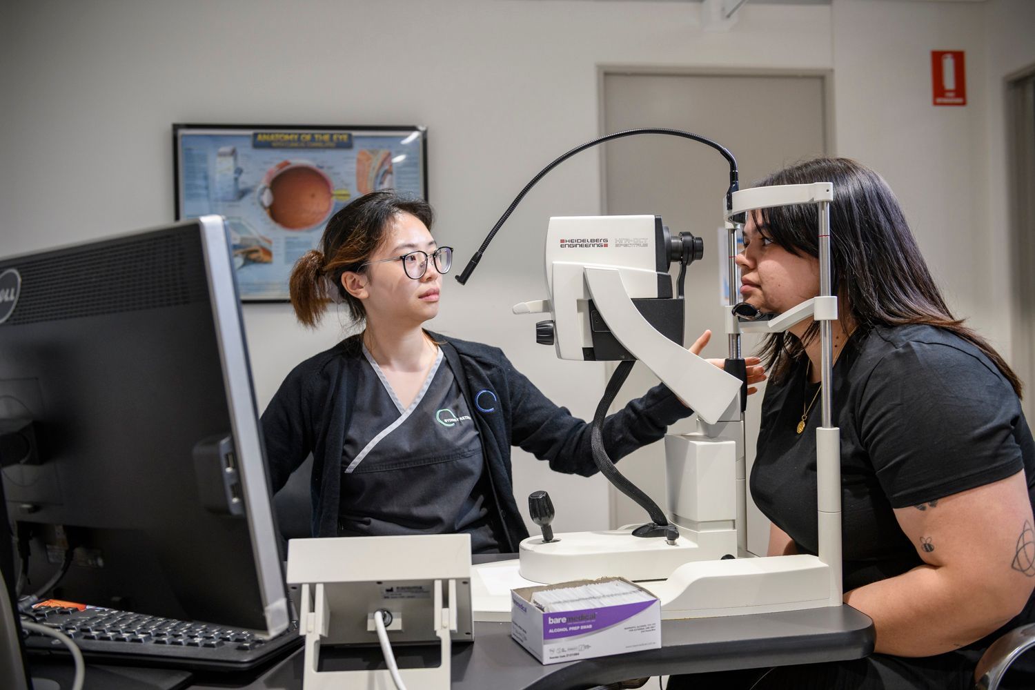
Retinal Vein Occlusion Diagnosis
Diagnosis typically involves fluorescein angiography, OCT scanning to assess macular swelling, and blood tests to identify underlying risk factors. If untreated, RVO may lead to serious complications such as macular oedema, neovascular glaucoma, vitreous haemorrhage, or retinal detachment.
Retinal Vein Occlusion Treatment Options
The blocked vein cannot be reopened. Treatments aim to protect vision and manage complications. These may include anti-VEGF injections, corticosteroid injections, or laser therapy. Vision outcomes vary, and ongoing monitoring is essential. The risk of RVO occurring in the other eye is approximately 5% per year.
Vision Outcomes After Retinal Vein Occlusion
Visual outcomes vary depending on how quickly treatment begins and whether the macula is affected. Some patients experience significant improvement with treatment, while others may have ongoing vision impairment. Early diagnosis and regular monitoring improve the likelihood of stabilising vision. Managing systemic health conditions plays an important role in long-term outcomes.
Permanent vision loss can occur in severe or untreated cases.
Retinal Vein Occlusion Frequently Asked Questions
Is retinal vein occlusion the same as a stroke in the eye?
It is often described this way, as it involves a blockage of blood flow, but the underlying mechanisms differ from a brain stroke.
Can retinal vein occlusion affect both eyes?
It usually affects one eye, but underlying risk factors may increase the chance of it occurring in the other eye.
Will my vision return to normal after treatment?
Some patients experience significant improvement, but vision may not fully return to normal depending on the severity.
Is retinal vein occlusion preventable?
Managing blood pressure, diabetes and cholesterol can help reduce risk, but it cannot always be prevented.
If you experience sudden vision changes or have been diagnosed with retinal vein occlusion, contact 02 9221 3755 to book an urgent assessment with our retinal specialists.